There are 5 major ways to image the body:
- CT
- X-ray
- MRI
- Ultrasound
- Nuclear medicine
- Scope
Other imaging modalities are simply variations on one of these 6 established ways to image the body. In this article we will review some of the major variations on these 6 techniques that you may come across in the hospital. Simple imaging like imaging of the chest or abdomen have been largely excluded from the list below.
At the end, we will have a short discuss on types of contrast.
CT
CT angiography
CT angiography is a procedure where contrast material is injected through a small catheter placed in a vein, followed by the use of a CT scanner to produce detailed images of arterial and venous blood vessels.
It is most useful to identify blood vessel abnormalities such as blood clots, arterial-venous malformation, aneurysms, dissections and stenoses.
What is the difference between a CT angiogram of the chest and a CT of the chest with IV contrast?
Both procedures involve a CT scan, and both involve the administration of IV contrast. The difference lies in the timing of the contrast and when the image is taken. A CT aniogram of the chest will timed so that the arteries or veins that we are interested in are filled with contrast. However with a CT of the chest with IV contrast, we presumably want to see the soft tissues and organs, so the contrast is timed so that more it is in the capillary beds between the arteries and veins.
CT enterography
CT enterography is a procedure where both IV and oral contrast material is provided to the patient followed by the use of a CT scanner to produce detailed images of both solid organs of the abdomen and the small bowel.
It is most useful to identify small bowel disorders such as Crohn’s disease, ulcerative colitis, Celiac disease, post-operative adhesions, and small bowel tumors.
CT colonography
CT colonography is considered a less invasive substitute for colonoscopy in patients who have comorbidities that prevent them from tolerating the sedation required in colonoscopy, or who have large masses that prevent the passage of the colonoscope through the rectum and colon. In CT colonography, first a tube or balloon is placed into the rectum so that air, typically CO2, can be pumped into the colon. After the colon has been sufficiently dilatated, CT images are taken with the patient in the supine and prone position.
Non-contrast CT head
In trauma patients or patients with signs and symptoms suggestive of a stroke, a non-contrast CT head is the first line of imaging. Non-contrast CT head is useful for ruling out subarachnoid hemorrhage, hematoma, midline shift, and skull fractures.
FDG PET/CT
PET scan is a procedure in which a radio-labeled glucose tracer called 18F-fluorodexoyglucose is administered to the patient before any imaging is performed. Highly metabolically active tissues use high levels of glucose, so areas such as the brain, areas of inflammation and especially cancer cells will take up a lot of this radio-labeled glucose tracer. Since the glucose is radio labelled as it decays it will release gamma rays which are detected by the PET scanner as dark areas on the images. The kidneys, ureters and bladder will also dark since FDG is renally excreted.
Unfortunately, the PET scan is not great for localization of specific structures, so often times a CT scan will be taken at the same time in order to determine specifically which anatomical regions of the body line up with the areas that light up during the PET scan.
CTPE/CTPA
CTPE (CT pulmonary embolism protocol) in many institutions is very similar if not the same as a CTPA (CT pulmonary angiogram). The main use of a CTPA is to detect a pulmonary embolism in high risk patients or low risk patients with a positive D-Dimer.
X-ray
Mammography
Mammography is simply the use of x-rays to screen for or diagnose breast cancer. Mammography is typically an unpleasant experience for most women since it involves the compression of the breast tissue along two planes in order to decrease unnecessary breast movements and reduce the dose of radiation required to visualize abnormalities. The 2 views acquired in a screening mammogram are the cranial caudal (top to bottom “birds eye”) view and the medial lateral oblique view (side view).
Radiologists will look for masses, calcifications, fibroglandular density, architectural distortion and asymmetry. Masses should be seen in 2 views. Radiologists can also comment on the degree of breast density. Breasts that are more dense have more fibroglandular tissue and are therefore more difficult to visualize (younger women have more dense breasts). Breasts that are less dense have more fat relative to fibroglandular tissue and are easier to image using mammography.
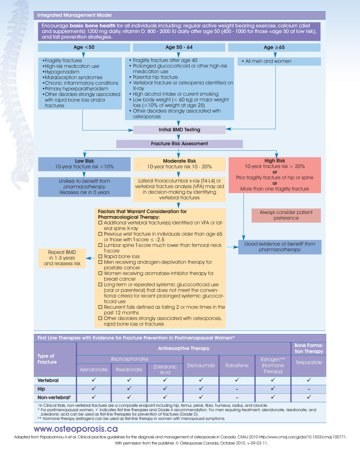
DEXA Bone scan
DEXA bone scan is a screening test for everyone over 65 (and younger based on specific criteria to look for signs of osteoporosis. The DEXA scan involves a central and peripheral examination. In the central DEXA examination, a specialized x-ray device measures bone density of the hip and spine whereas the finger, hand, forearm or foot is assessed in the peripheral DEXA examination.
After the test is done a T score and Z score will be reported. The T score is reported as the number of standard deviations above or below the patient’s bone density as compared to what is expected for a healthy and young person the same sex.
- Normal – T-score > -1
- Osteopenia – T-score between -1 and -2.5
- Osteoporosis – T-score equal to or less than -2.5
The Z-score is reported as the number of standard deviations above or below the patient’s bone density as compared to what is expected for someone of the patient’s age, sex, weight, and ethnic or racial origin. A Z-score less than -1.5 should raise suspicion for an underlying cause of osteoporosis that isn’t simply due to natural aging (as most elderly patients have osteoporosis, but the Z score includes age so if they still have a low Z score then there may be some underlying cause).
For more information on osteoporosis vs osteomalacia check out this article!
Acute abdominal series
The acute abdominal series is a series of radiographs that are ordered in patients with acute abdominal pain. There are 4 main views
- Supine view
- General overview of abdomen
- Bowel gas pattern
- Calcifications
- Masses
- Prone (or lateral rectum) view
- Useful to see if there is gas in the rectum or sigmoid (the highest points of the large bowel when prone)
- Useful to see if there is gas in the ascending and descending colon
- Lateral rectum is a substitute view which involves the patient lying on their left side with both their legs partially flexed
- Upright (or left lateral decubitus) view of abdomen
- Useful for free air in peritoneum
- Useful to detect air fluid levels within the bowel lumen
- Left lateral decubitus is when the patient lies on their left side
- Upright (or supine ) view of chest
- Useful for free air under the diaphragm
- Detection of acute abdomen mimics
- Pneumonia
- Pleural effusions (which in itself can be secondary to abdominal abscesses, pancreatitis and ovarian tumors)
KUB
KUB stands for kidneys, ureters and bladder and is the equivalent of a supine abdominal radiograph (although you can also order additional views of KUB). It is most useful for detecting urinary tract stones, appropriate tube placement and bowel distention. However, since the patient is supine, you cannot see air-fluid levels (so you cannot rule out free air) and you also cannot rule out constipation using a KUB.
Fluoroscopy
Fluoroscopy is basically when you take many x-rays in rapid succession in order to produce a video of continuous and live visualization of the body. Fluoroscopy is basically a live x-ray. Because it requires multiple rapid x-rays, often paired with radiocontrast, there is a significant increase in radiation compared to a normal plain film x-ray.

Barium studies
CT, ultrasound and MRI have largely replaced the use of barium studies, but under certain indications they are still performed.
Barium studies are rarely indicated for acute pain!
Modified Barium Swallow (MBS) also known as videofluoroscopic swallowing study (VFSS)
A modified barium swallow study refers to the assessment of dysphagia by speech language pathology. In a MBS, the patient is asked to swallow a thick barium mixture as well as barium laden foods, while real time fluoroscopic images are being taken. The SLP can then look at the video to see whether the patient is at risk for aspiration or assess for dysphagia. The MBS assesses the area from the mouth to the hypopharynx.
Barium Swallow (BaSW) or esophagram
A barium swallow is similar to a MBS except it extends beyond the hypopharynx to study the more distal esophagus – specifically from the hypopharynx to the proximal stomach. While it may be performed for dysphagia (MBS is usually done first), it can also be performed if one suspects a esophageal mass, dysmotility or esophageal stricture. It can be performed with double or single contrast.


Upper GI study (UGI)
An upper GI study is a fluoroscopic procedure that studies the area from the esophagus to the ligament of treitz. The ligament of treiz is also called the suspensory muscle of the duodenum. It attaches from the dudodenojejunal junction to the left crus of the diaphragm. The ligament of treitz is also considered to the division point between upper and lower GI bleeds. The right and left crus are the tendons that attach the diaphragm to the vertebral column.
It is used in cases of epigastric pain, post-gastric or duodenal procedure or surgery, gastritis, duodenitis, gastric outlet obstruction, suspected midgut volvulus in infants or ulcer. It can be performed with double or single contrast. In double contrast, in addition to drinking the barium contrast, patients are also given baking soda crystals which release gas and dilate the upper GI tract.
UGI is the investigation of choice for suspected midgut volvulus in infants!
An upper GI series with oral contrast is the standard for any infant with new onset acute bilious emesis!
Small bowel follow through (SBFT)
Small bowel follow through is a fluoroscopic procedure that studies the area from the duodenum to the ileocecal valve (the junction between the ileum and the cecum of the colon). It also involves the patient drinking barium contrast. As it passes through the small intestine, fluoroscopy is used to look for any abnormalities in size and shape of the small intestine. Small bowel follow through is called that in part because it usually follows an UGI fluoroscopic study as discussed above, although it can be performed without UGI.
Small bowel follow through is best used to assess for abnormalities in the size and shape of the small intestine. While it may be used in evaluating Crohn’s disease since Crohn’s affects the GI tract from gum to bum, if CT or MR enterography is available that should be performed first.
It is used in cases of Crohn’s disease, small bowel obstruction, masses, cancer or poylps in the small intestine, post-abdominal or bowel surgery if suspected complications are present.
Barium Enema (BE) or Lower GI study (LGI)
Barium enema is a fluoroscopic procedure which involves the instillation of barium contrast into the rectum and throughout the large colon. It studies the area from the cecum to rectum.
It is used to diagnose benign polyp tumors, cancer, ulcerative colitis, and Hirschsprung disease in children.
It can be performed with single or double contrast. In double contrast it is much more important the patient has a clean colon, compared to single contrast barium enema.
Avoid barium enemas in inflamed and friable colons as it increases the risk of perforation!
Coronary catheterization (also known as coronary angiography) and PCI
In patients who are suspected to have a STEMI or severe NSTEMI, fluoroscopy can be used to visualize and identify stenosed or completely occluded coronary arteries.
A needle is inserted into the femoral artery, followed by the passage of a wire through the needle, that is guided through the arteries and eventually reaches the heart. Next the catheter is place over the guide wire, and passes through the arterial system to also reach the heart and its tip is placed within the lumen of one of the coronary arteries. The catheter is intentionally designed to be have a smaller diameter than the lumen of the artery in which it will be placed. Placement of the catheter is performed under fluoroscopy guidance.
Once the catheter is in the coronary artery (but not occluding the coronary artery), the guide wire is removed as it is no longer needed. Next the radiocontrast is injected into the catheter which is delivered into the coronary arteries where fluoroscopy can now be used to evaluate for any occlusion or stenoses of the coronary arteries. With each cardiac cycle the radiocontrast dye is washed away, so it has to be repeatedly administered.
If an occluded or stenosed coronary artery is found on coronary angiography, then PCI (percutaneous coronary intervention) is performed which refers to the placement of a drug eluting or metal stent in the occluded or stenosed coronary artery.
PICC Line Insertion
Fluoroscopy is a great tool that can be used for the insertion of a catheter into any vein or artery in the body. A common use for it is for the insertion of PICC lines. A PICC line is a catheter that is inserted into a peripheral vein and coarsed until it reaches the right atrium or SVC. PICC lines can also be performed under ultrasound guidance as well.
For more information on PICC lines and other central lines, check out our article on central and arterial lines!
Spine and joint injections and lumbar puncture
In patients with spinal deformities such as scoliosis, it may be difficult to perform a lumbar puncture without imaging or with ultrasound. In these unique circumstances, fluoroscopy guided lumbar puncture may be used.
Injections into the joint or spine can also be performed under fluoroscopic guidance.
MRI
MR Angiography (MRA)
MRI angiography (like CT angiography) is a procedure where gadolinium contrast is injected through a small catheter placed in a vein, followed by the use of MRI to produce detailed images of arterial and venous blood vessels. Gadolinium is much less likely than iodine contrast to induce a allergic reaction.
It is most useful to identify blood vessel abnormalities such as blood clots, arterial-venous malformation, aneurysms, dissections and stenoses.
MR Enterography
MRI enterography is a procedure where both IV and oral contrast (gadolinium) material is provided to the patient followed by the use of MRI to produce detailed images of both solid organs of the abdomen and the small bowel.
It is most useful to identify small bowel disorders such as Crohn’s disease, ulcerative colitis, Celiac disease, post-operative adhesions, and small bowel tumors.
MRI Breast
Breast MRI is not routinely performed but is still increasingly being used for patients with known breast cancer to screen for additional foci of breast cancer within the affected and contralateral breast, especially given how much more favored lumpectomy is over a total mastectomy. Other reasons for a breast MRI is to screen for breast cancer in high risk patients (BRCA carriers, strong family history) and for evaluation of breast implants. MRI however does not replace mammography in patients of any risk status, but rather can supplement breast ultrasound and mammogram.
fMRI
fMRI stands for functional MRI and through detecting changes in blood flow that vary in brain activity it is able to determine which areas of the brain are active during certain activities. For instance, the radiologist or technician may ask the patient to read, speak, listen to music or speech and then look at which areas of the brain “light up” so to speak.
It is used in cases of stroke, trauma or degenerative disease and to evaluate the functional anatomy of the brain. It is also used in the planning for major neurological surgery.
Ultrasound
Transvaginal vs Transabdominal ultrasound
Transvaginal ultrasound involves placing the ultrasound probe through the vaginal canal in order to get a better view of the endometrium and adnexa (that usually cannot be obtain via transabdominal ultrasound).
Transvaginal ultrasound is commonly used to detect
- Fibroids
- Polyps
- Uterine cancer
- Ovarian cysts
- Ectopic pregnancy
Transabdominal ultrasound can be used to visualize most of the abdominal organs including the liver, gallbladder, kidneys, pancreas, appendix, bladder, uterus, ovaries, spleen, stomach, aorta, and IVC.
Common/important uses and its corresponding organ
- Gallbladder – cholecystits, cholangitis
- Aorta – aortic dissection, aortic aneurysm
- Adnexa – ectopic pregnancy
- Kidneys – incidental rising creatinine, kidney injury, hydroureter
- Appendix – appendicitis
In ectopic pregnancy the decision of whether to use a transabdominal or transvaginal ultrasound depends on the level of bHCG. If the quantitative bHCG is greater than 5000 mIU/mL then a normal intrauterine pregnancy should be detectable on transabdominal ultrasound. If the quantitative bHCG is greater than 1500 mIU/mL then a normal intrauterine pregnancy should be detectable on transvaginal ultrasound. If either of these is not true and you cannot detect a pregnancy despite the corresponding levels of bHCG, this suggests an ectopic pregnancy!
Ultrasound breast
Ultrasound is another modality in addition to mammogram to detect breast cancer. It is commonly used in women with especially fibrous breast tissue where detecting a malignancy using mammogram poses a challenge. It never replaces mammography but can supplement it. Ultrasound can also be used in pregnant women instead of mammography.
Echocardiogram
A form of ultrasound that specifically looks at the heart. There are 2 main types of echocardiogram: transthoracic echocardiogram and transesophageal echocardiogram.
Echocardiogram is used to detect wall motion abnormalities, to estimate ejection fraction in patients with heart failure, to look for valvular abnormalities, to look for valve vegetations in patients with Staphlococcus aureus bacteremia or suspected endocarditis, and to look for an intracardiac thrombus or patent foramen ovale in patients in stroke or atrial fibrillation (without anticoagulation).
Nuclear medicine
Bone scan (skeletal scintigraphy)
Bone scans is a nuclear medicine procedure that involves the injection of the radiotracer 99mTc with methylene diphosphonate (MDP). Bone is composed of organic component osteoid and inorganic component hydroxyapatite (which in turn is composed of Ca and PO4). The MDP when near bone will adsorb onto the hydroxyapatite of the bone. Since mineralization occurs where there is bone growth, the areas of bone with more turnover and (and therefore more mineralization) will have more 99mTc with methylene diphosphonate adsorbed to it. MDPs job is done at this point. Next the Tc which is the radioactive part of this compound will give off gamma rays as it decays which is then detected by a special gamma ray camera.
It is useful in the diagnosis of osteomyelitis (differentiating cellulitis from osteomyelitis), difficult fractures that don’t show up on x-ray and cancer (that may have metastasized to bone).
Scope
Esophagogastroduodenoscopy (upper GI endoscopy)
Esophagogastroduodenoscopy also known as EGD is a procedure involving placing a scope through the mouth all the way through to the duodenum to directly visualize (and biopsy if necessary) the upper GI tract. It can be used to diagnose
- Esophageal varices
- Upper GI bleed
- Crohn’s disease
- Celiac disease
- Peptic ulcer
- Tumors
- Esophageal stricture
Colonoscopy and flexible sigmoidoscopy
Colonoscopy is the same as an EGD, except it looks at the lower GI tract (the entire colon until the cecum). Flexible sigmoidoscopy is similar to a colonoscopy except it only goes as far as the splenic flexure.
They are both used for lower GI bleeding, screening or diagnosing colon cancer and the detection of polyps or IBD.
